



Home About Curriculum Workflow Reviews Articles FAQ
DIGITAL ORTH MASTERS ACADEMY

MASTER DIGITAL ORTHODONTICS

From Scan · To · Smile

Not ready yet? Start with the free course

SCROLL
Dr. Mohamed Hesham — Digital Orth Masters
10+
Years Clinical Experience

Dr. Mohamed
Hesham

Pioneer of digital orthodontics in the Arab world. Founder of the H Digital Anchor TADs System and DJED Digital Orthodontics LAB — Dr. Hesham has redefined what's possible in orthodontic treatment planning.

  • BDS — Alexandria University, 2014
  • Morth Residency — Royal Colleges of Surgeons, Edinburgh
  • CEO — Hesham Dental House Clinic
  • Founder — DJED Digital Orthodontics LAB
  • Founder — H Digital Anchor TADs System
  • Founder — Digital Orth Masters Academy
VIEW CURRICULUM →

The Future of
Orthodontic Education

01
🔩

PURE SKELETAL ANCHORAGE

Master TADs placement with CBCT-guided surgical planning. Eliminate anchorage loss permanently from your clinical practice.

02

48-HOUR LAB TURNAROUND

Design and deliver digital appliances faster than ever. Cut lab dependency and unlock a new revenue stream per case.

03
🎯

HIGH SUCCESS RATE

Mini screws placed with precision in the palate using digital surgical guides. Documented high success rates across complex cases.

04
💰

REDUCE COSTS 50%

Design your own appliances digitally. Sell them as a separate service before braces — independent pricing, maximum value.

05
🖥️

ONYXCEPH3™ MASTERY

Full license included. Master the world's most advanced orthodontic digital planning software with unlimited server access.

06
🌍

GLOBAL COMMUNITY

Join orthodontists from Iraq, Algeria, Morocco, Egypt and beyond. Continued shadowing & mentoring via TeamViewer after the course.

Course Curriculum

DAY 01
Day 1

INTRODUCTION TO DIGITAL ORTHODONTICS

  • Limitation of conventional methods
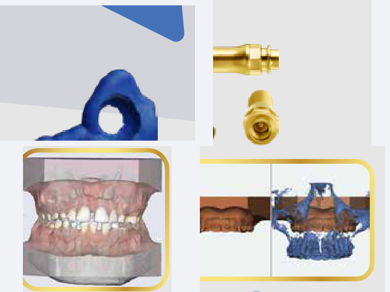
  • Scanning & 3D Printing workflow
  • OnyxCeph3™ Data Entry & CBCT
  • TADs Design, First vs Last, Uses
DAY 02
Day 2

TADs IN ONYXCEPH & SURGICAL GUIDE

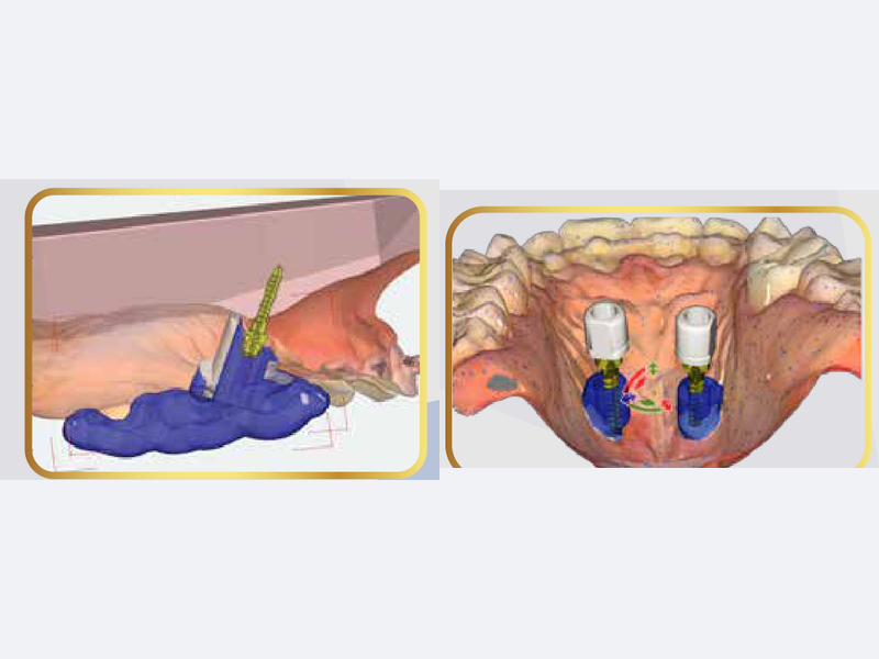
  • TADs Placement on OnyxCeph
  • Surgical Guide Fabrication
  • 2 Screws vs 4 Screws protocol
  • Basic Aligners (NEW)
DAY 03
Day 3

MARPE & SKELETAL EXPANSION

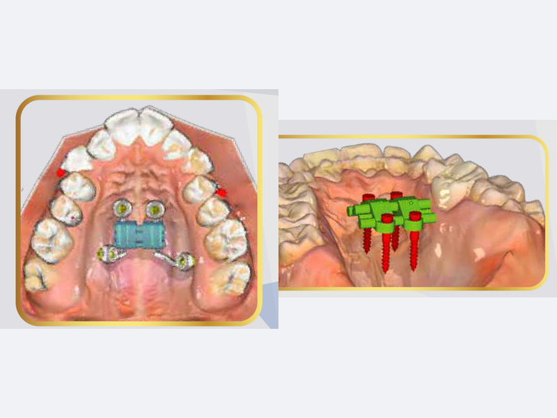
  • MARPE Biomechanics & Application
  • MARPE Appliance Design
  • MSE Design & Limitation
  • Guided MSE Protocol
DAY 04
Day 4

PART 1ADVANCED DIGITAL APPLIANCE DESIGN

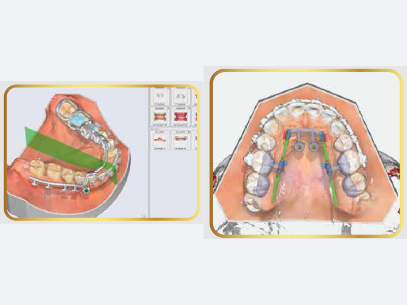
  • Mouth Trap (Intrusion)
  • Distalizer
  • Mesaliser
DAY 05
Day 5

PART 2ADVANCED DIGITAL APPLIANCE DESIGN

  • Disimpaction Appliance
  • Canine First Appliance
  • Twin Blocks
  • Mara Appliance
DAY 06
Day 6

PART 3ADVANCED DIGITAL APPLIANCE DESIGN

  • Appliance Bonding & Delivery
  • Full LAB Procedures

From Scan to Execution

🦷
1
INTRAORAL SCAN
Capture full arch digital impression
📡
2
CBCT ANALYSIS
3D bone mapping for precision planning
🎯
3
TADs PLANNING
Digital TAD placement & surgical guide
⚙️
4
APPLIANCE DESIGN
Full digital appliance in OnyxCeph3™
🖨️
5
3D PRINT & DELIVER
48-hour lab return, precision execution
// SCAN QUALITY
ACCURACY
COVERAGE
RESOLUTION
// TAD PLACEMENT
BONE DENSITY OK ✓
SAFE ZONE CLEAR ✓
ROOT CLEARANCE ✓
// APPLIANCE OUTPUT
DESIGN TIME
PRINT READY
DELIVERY SPEED

Hear from Our Happy Learners

F-Ortho Residency
Dr. Abdullah Mohamed
F-Ortho Residency
Dr. Saeed Ibrahim
F-Ortho Instructor
Dr. Asmaa Ebada
F-Ortho Residency
Algerian Orthodontist

Real Results

★★★★★

شكراً جزيلاً على الكورس والسلاسة — فعلاً خليت السوفتوير يبين سهل ويارب يكمل كدا 🙏

🇩🇿
Orthodontist
Algeria
★★★★★

I am really excited for the next module. I learned a lot from you — the most comprehensive digital ortho course I've attended.

🇮🇶
Dr. Ibrahim
Iraq
★★★★★

Thank you for the explanation — it was a very interesting course last week. Salam Dr Hesham! 🇲🇦

🇲🇦
Dr. Essalhi Assia
Morocco
★★★★★

الكورس تحفة وتسلم جداً — شرح وافي وكافي ومفصل بكل الخطوات. والله الواحد محسسش بالوقت من حلاوة الكورس.

🇪🇬
Islam Sameh
Egypt
★★★★★

الدكتور اصلاً ما مقصر — رهيب ربنا يزيده علم ومعرفة ❤️

🇮🇶
Dr. Elaphharb
Baghdad, Iraq
★★★★★

الصراحة كورس غاية في الجمال — الد محمد هشام رائع بكل المقاييس والتنظيم وكل حاجة تحفة حرفياً ماشاء الله 🙏

🇪🇬
Orthodontist
Egypt

Your Certification

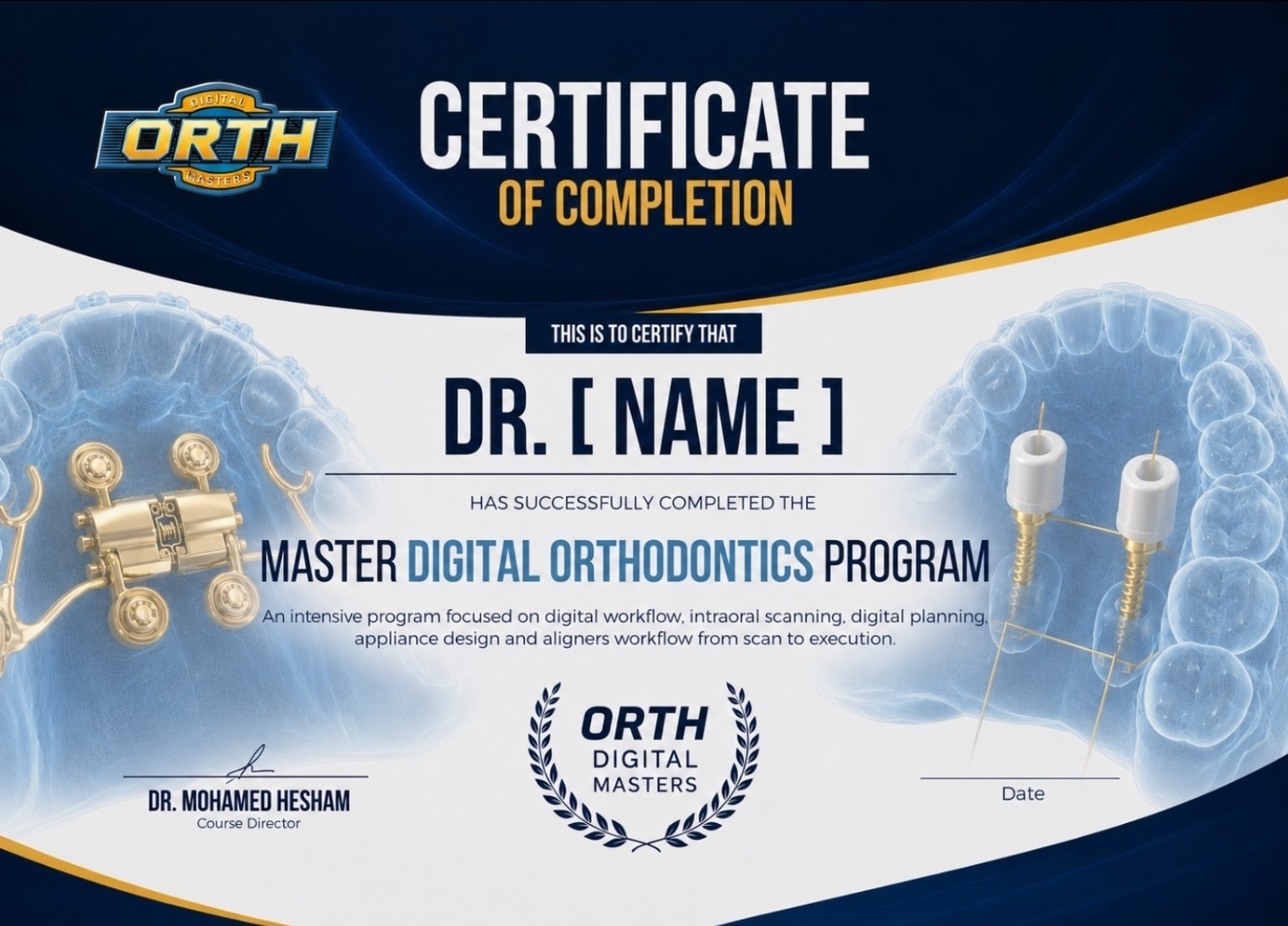
Digital Orth Masters Certificate of Completion
DOM CERTIFIED

CERTIFICATE OF COMPLETION

Digital Orth Masters Certificate

Upon successful completion of the 6-day intensive program, you receive an official Digital Orth Masters certification — recognizing your mastery of digital orthodontic workflow.

  • Official DOM Academy Certificate
  • Full OnyxCeph3™ License Included
  • Access to all 3D appliance STL templates
  • 6 months recorded course access
  • TeamViewer shadowing & mentoring
  • Money back guaranteed within 7 days

Latest Articles on The Blog

FAQ

Yes — every session is fully recorded and stays available for 6 months from your enrolment date, so you can rewatch any module whenever it suits you.
This program is built for orthodontic specialists, or for practitioners with at least two years of hands-on orthodontic cases. The content is advanced — it does not cover the basics.
Not at all. There is a fully online track that covers the workshop day and the live demo as well — and an offline track if you would rather attend in person.
Your lab bill drops by up to 50%, your patient gets treated faster, and you stay in full control of every detail of the digital appliance.
Absolutely. Pay a deposit to reserve your seat, then settle the remaining balance after the first lecture — no pressure.
FREE ACCESS

Still deciding?

Get a taste of the digital workflow first. Watch the free introductory course — no payment, no commitment.

GET THE FREE COURSE

READY TO MASTER
DIGITAL ORTHODONTICS?

Join hundreds of orthodontists who transformed their practice with the Digital Orth Masters program.

Or start with the free course if you want to see the workflow first.

Blog / Research & Sources
SEMINARS IN ORTHODONTICS · 2024 · VOL 30

Kilbon Appliance

Antero-posterior lingual retractor design and biomechanics

Why This Matters

Anterior open bite in an adult is one of the least forgiving problems in orthodontics. It has no single cause — genetics, skeletal pattern, tongue and soft-tissue behaviour, and long-standing habits all feed into it — and whatever you do to close it, relapse is always waiting. When the same patient also presents with lip protrusion and excessive incisor or gingival display, the treatment problem multiplies: you now need to retract the anterior segment, intrude it, and intrude the posterior segment, all at once, without the appliance itself becoming the patient’s next complaint.

The conventional answer has been surgery, or labial mechanics with buccal miniscrews. This paper proposes a third route: an antero-posterior lingual retractor (APLR) anchored to TADs in the midpalatal area, capable of retracting the anterior teeth and intruding the entire maxillary dentition simultaneously — while staying completely invisible from the front. The authors lay out the design, the force system behind it, and three treated cases showing how the same appliance behaves differently depending on lever-arm length, TAD height, tube angulation, and palatal vault depth.

The Problem With Labial Total-Intrusion Mechanics

Before getting to the APLR, the paper works through why the existing options are unsatisfying. In a patient with an open bite and a gummy smile, intruding only the molars is not enough — the whole maxillary dentition needs to come up, which produces something close to a total maxillary impaction and lets the mandible autorotate counterclockwise. That autorotation is the mechanism that closes the bite and brings the chin forward.

Earlier work achieved this with two miniscrews per buccal side, positioning the appliance so that the centre of resistance of the maxillary dentition sits between the two lines of force. Other groups used a transpalatal arch to stop the molar crowns from tipping buccally toward the screws, or an archwire with an accentuated curve of Spee combined with buccal TADs to cancel the unwanted extrusive component in the premolar region.

All of these work. All of them share two drawbacks: the hardware is highly visible at exactly the moment a protrusive, gummy-smile patient is most self-conscious about their smile, and anatomy in the buccal alveolus limits both how high you can place the TAD and how long you can make the lever arm — the two variables that actually govern the force vector.
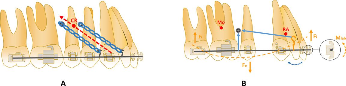
Figure 1
FIG. 1 — Labial approaches to total maxillary intrusion. A: two buccal miniscrews per side, used for intrusion and distalisation in a non-extraction case. B: a single buccal TAD per side combined with an accentuated curve of Spee for intrusion plus anterior retraction in a premolar extraction case.

Where Conventional Lingual Retractors Fall Short

A lingual retractor bonds attachment pads to the palatal surfaces of the anterior teeth, joined by a lingual arch into a single anterior splint. Lever arms of varying length are soldered to that splint, and elastic chain or coil springs run from the lever arms back to TADs in the midpalatal area or on the palatal slope.

The paper makes a good case for why this is attractive beyond aesthetics. First, retraction can start immediately — you are not stuck doing the usual align-and-level-then-retract sequence that flares the incisors labially before it brings them back, so round-tripping is minimised and the patient sees their chief complaint improving early. Second, friction is almost absent: the only metal-on-metal contact is between the guide bar and the posterior tube, so extraction space closes far more efficiently than it does sliding brackets along an archwire. Third, and most importantly, you can steer the force vector precisely by changing lever-arm length and TAD position.

The limitation is what happens when that vector is wrong. With the C-lingual retractor, if the line of action passes below the centre of resistance of the anterior segment, the segment rotates clockwise — you lose anterior torque and open a canine bite. The Double J retractor added torque springs on the lever arms to fight this. But both are anchored only to the anterior teeth. Neither can intrude the posterior segment, which is precisely what an open-bite, gummy-smile patient needs.
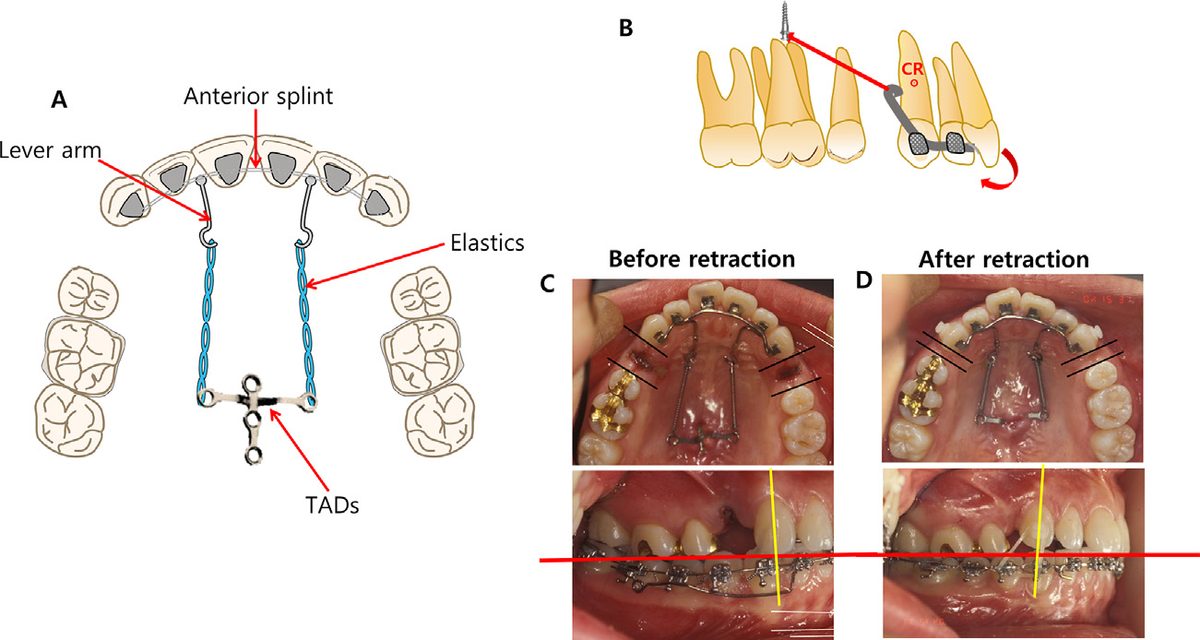
Figure 2
FIG. 2 — The C-lingual retractor and its failure mode. A: appliance schematic. B: line of action passing below the centre of resistance of the anterior teeth. C–D: the resulting clockwise rotation of the anterior segment — torque loss and canine open bite — before and after retraction.

How the APLR Is Built

The single structural difference between the APLR and a conventional lingual retractor is that the APLR also grips the posterior teeth. That one change is what converts it from an anterior-retraction device into a whole-arch intrusion device.

  • The anterior segment is essentially unchanged: pads on the palatal surfaces of the anterior teeth, joined into a splint, with lever arms soldered on.
  • 0.036-in stainless steel guide bars are soldered to the lever arm or the front attachment pad and run posteriorly, passing through a tube on the posterior segment.
  • A posterior splint is bonded to the palatal surfaces of the second premolar and the first and second molars, reinforcing the anchor unit.
  • A posterior tube is soldered at the first molar for the guide bar to slide through. Two tubes can be added per side if a TPA is being used.
  • If the posterior teeth themselves need intrusion or torque control, a hook can be soldered to the TPA.
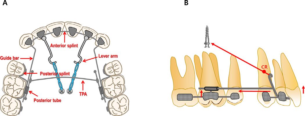
Figure 3
FIG. 3 — The APLR. A: component schematic showing the anterior splint, lever arms, guide bars, posterior splint, posterior tube and TPA. B: the resulting force system when elastics run from the lever arm to a midpalatal TAD.

The Biomechanics — What Each Variable Actually Controls

This is the most clinically useful part of the paper. Rather than presenting the APLR as a fixed recipe, the authors treat it as a set of dials, each with a predictable effect:

  • Lever-arm length and TAD height together set the line of action. If that line passes under the centre of resistance of the anterior teeth, you get retraction with tipping. If it passes through the CR, you get bodily movement. Bodily retraction therefore demands longer lever arms and TADs placed higher.
  • In hyperdivergent Class II patients with open bite and a gummy smile, TADs must sit higher than the lever-arm hook — otherwise the total intrusion component simply is not generated.
  • The guide bar controls the retraction vector and protects anterior torque. Because the bar is soldered to the anterior splint and constrained by the posterior tube, it prevents the anterior segment from rotating as it retracts.
  • Posterior tube angulation is the intrusion dial. A tube parallel to the occlusal plane produces bodily movement of the anterior teeth; tipping the tube distally increases the amount of anterior intrusion. This is the cheapest, most direct way to tune vertical control in gummy-smile cases.
  • Non-parallel force and guide bar generates posterior intrusion. When the APLR force direction and the guide bar are not parallel, an intrusive component appears in the posterior segment — and total intrusion of the maxillary dentition is what rotates the mandible counterclockwise and closes the open bite.

The Posterior Segment Has to Behave as One Unit

The paper is emphatic about a failure mode that is easy to overlook. If the posterior dentition is not splinted into a single unit, the intrusive force is delivered only to the first molar — the one tooth the posterior tube happens to be bonded to. That tooth intrudes in isolation and everything else stays put.

Three acceptable ways to connect the posterior segment are described: bonding buccal brackets or tubes from the canine back and engaging a heavy rectangular segmental archwire; bonding a posterior splint to the buccal surfaces; or bonding a posterior splint to the palatal surfaces. There is also a related caution — because the intrusive load reaches the posterior teeth only through their palatal surfaces, some palatal tipping is expected, and a TPA is the standard countermeasure.
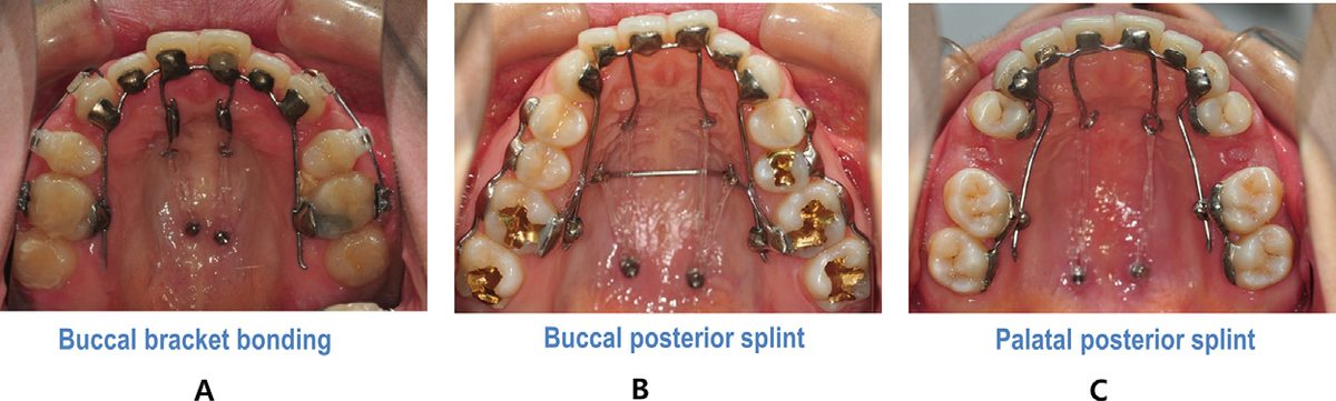
Figure 5
FIG. 5 — Three validated ways to splint the posterior dentition into a single unit. A: buccal bracket bonding from canine to molars with a heavy segmental rectangular archwire. B: buccal posterior splint. C: palatal posterior splint.

The Mandibular Molars Can Undo Your Work

One more mechanism deserves attention, because it quietly limits how much you get back for the intrusion you achieve. As the maxillary dentition intrudes, the mandibular molars have room to extrude compensatorily — and every millimetre of that compensatory extrusion cancels part of the mandibular autorotation you were counting on. The risk is highest exactly where these patients live: steep occlusal planes and high mandibular plane angles. If you expect it, plan for it — mandibular posterior TADs are the straightforward answer. The three cases below show this playing out very clearly.

Case 1 — Long Lever Arms, Deep Palatal Vault

A 25-year-old woman presented with lip protrusion and anterior open bite, a convex profile with a retrusive chin, a gummy smile and a flat smile arc. Cephalometrics showed a skeletal Class II hyperdivergent pattern (ANB 7.2°, SN-GoMe 42.9°) with normally inclined maxillary incisors (U1-FH 114.1°) and proclined mandibular incisors (IMPA 104.1°). She specifically requested a less visible appliance and early improvement of her protrusion.

Treatment involved maxillary first premolar extraction plus removal of poor-condition mandibular first molars and a supernumerary tooth. Because bodily retraction was wanted, roughly 15 mm lever arms were soldered to both canine pads and the posterior tube was tipped distally to boost anterior intrusion. TADs went into the midpalatal suture; the patient’s deep palatal vault meant the posterosuperior force vector produced a strong intrusive component. Space closure was essentially complete twelve months after APLR delivery; total treatment ran 28 months.

Outcome: lip protrusion, open bite and gummy smile all fully resolved with a consonant smile arc. Superimposition showed relatively bodily retraction (U1-FH 112°), 1.0 mm of maxillary anterior intrusion and 2.5 mm of maxillary molar intrusion. But the mandibular molars extruded 1.5 mm compensatorily — so counterclockwise rotation fell short of expectation (SN-GoMe 42.1°) and ANB barely moved (6.8°).
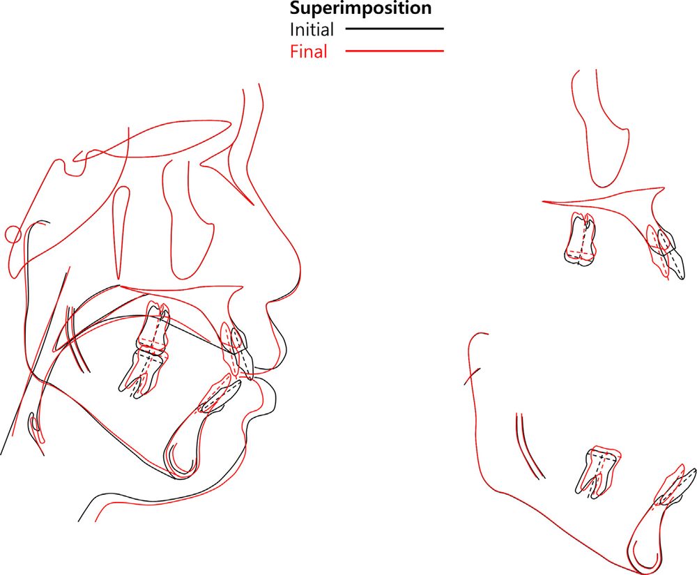
Figure 8
FIG. 8 — Case 1 — superimposition of lateral cephalograms before (black) and after (red) treatment. Anterior retraction and maxillary intrusion are evident, but compensatory extrusion of the mandibular molars limited the expected counterclockwise rotation.

Case 2 — Short Lever Arms, and the Difference Mandibular TADs Make

A 24-year-old man with lip protrusion and AOB, a retrognathic mandible, gummy smile, lip incompetency and moderate crowding. Skeletal Class II hyperdivergent, notably more severe vertically than Case 1 (ANB 6.3°, SN-GoMe 52.7°), with proclined incisors in both arches and bilateral condylar resorption visible on the panoramic radiograph. Surgery was offered as the primary option and declined.

Maxillary second premolars and mandibular first premolars were extracted. Here short lever arms were used, deliberately setting the line of force below the CR to retract with controlled tipping. Posterior tubes were again tipped distally, a TPA guarded against palatal tipping, and midpalatal TADs provided anchorage. Critically, additional TADs were placed between the mandibular first and second molars — both to help retract the mandibular incisors and specifically to block the compensatory extrusion that had limited Case 1. Power chain between the midpalatal TADs and the TPA added extra molar intrusion. Total treatment: 29 months.

The contrast with Case 1 is the point of the case. Retraction with controlled tipping (U1-FH 107.1°), 1.5 mm anterior and 2.5 mm molar intrusion — but no compensatory extrusion of the mandibular molars. The mandible rotated significantly counterclockwise (SN-GoMe 50.1°) and ANB dropped to 5.0°. Despite the history of degenerative arthritis, no further condylar resorption occurred.
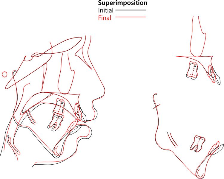
Figure 11
FIG. 11 — Case 2 — superimposition before and after treatment. With mandibular molar extrusion actively blocked by TADs, the mandible rotated counterclockwise as intended and ANB reduced meaningfully.

Case 3 — When the Palatal Vault Is Shallow

A female patient with lip protrusion and AOB, convex profile, retrusive chin, lip incompetency, and 4–5 mm lip protrusion relative to the E-line. Skeletal Class II hyperdivergent (ANB 7.0°, SN-GoMe 43.3°), normally inclined maxillary incisors (U1-FH 118.0°), proclined mandibular incisors (IMPA 97.8°).

Maxillary first premolars, third molars and mandibular second premolars were extracted for a Class I molar relationship. The APLR was bonded after aligning the six maxillary anterior teeth. Bodily retraction was the goal, so long lever arms of about 17 mm were soldered between the central and lateral incisors so the force would pass close to the CR. Because less anterior intrusion was needed here, the posterior tube was left parallel to the occlusal plane rather than tipped distally — a direct demonstration of the tube-angulation dial being used in the opposite direction from Case 1. Treatment ran 27 months.

Results: protrusion and open bite fully resolved, midline in harmony with the facial midline, improved profile and mentalis strain. Superimposition showed relatively bodily retraction (U1-FH 113.0°) with only mild intrusion — 0.5 mm anterior, 1.5 mm molar — and 0.5 mm of compensatory mandibular molar extrusion. Counterclockwise rotation was slight (SN-GoMe 42.5°), producing an anterosuperior shift of menton and a small reduction in lower anterior facial height, with ANB essentially unchanged at 6.3°. The authors are explicit that greater autorotation would have required actively controlling mandibular first molar extrusion. Her shallow palatal vault is offered as the reason the intrusive force stayed relatively small — anatomy, not technique, set the ceiling.
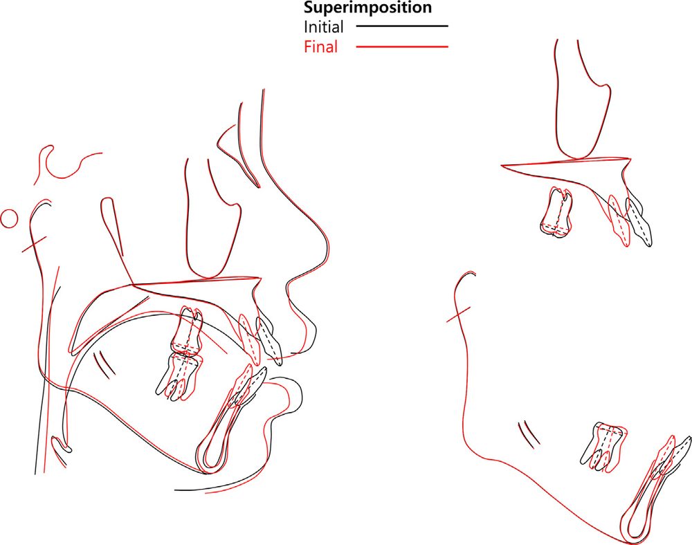
Figure 14
FIG. 14 — Case 3 — superimposition before and after treatment. Bodily retraction was achieved, but with a shallow palatal vault and no mandibular molar control, vertical change and autorotation were modest.

Key Findings

  • The APLR does two jobs at once. Anterior retraction and total maxillary intrusion from a single invisible appliance, in patients who would otherwise be candidates for surgery or highly visible labial mechanics.
  • Lever-arm length plus TAD height decide tipping vs bodily movement. Line of action under the CR → controlled tipping; through the CR → bodily retraction, which needs longer arms and higher TADs.
  • Posterior tube angulation is the vertical-control dial. Parallel to the occlusal plane for bodily movement; tipped distally for more anterior intrusion.
  • The posterior segment must be splinted as one unit — otherwise intrusive force is wasted on the single molar carrying the tube. A TPA should accompany it to limit palatal tipping.
  • Palatal vault depth caps what is achievable. Deep vaults allowed a stronger intrusive vector (Cases 1 and 2); the shallow vault in Case 3 produced markedly less intrusion for comparable mechanics.
  • Compensatory mandibular molar extrusion is the main thief of autorotation. Case 2 — the only case with mandibular posterior TADs — was the only one with no compensatory extrusion, and the only one with meaningful counterclockwise rotation and ANB reduction.
  • Friction is minimal and round-tripping is avoided, since retraction begins immediately rather than after conventional levelling.
Clinical takeaway: Across all three cases the maxillary mechanics performed as designed — anterior retraction and maxillary intrusion were achieved every time. What varied was how much of that intrusion translated into mandibular autorotation, and the deciding variable was not the maxillary appliance at all. It was whether the mandibular molars were prevented from extruding, and how deep the palatal vault was. Plan the mandibular arch and read the vault before you commit to a vertical target.

What This Means for Practice

For a digital workflow, the value here is that every APLR variable the authors identify — lever-arm length, TAD position and height, guide-bar path, posterior tube angulation, splint extent — is a design parameter that can be planned on a virtual setup and CBCT before anything is soldered. This appliance is unusually well suited to CAD design and digital transfer precisely because its behaviour is so sensitive to a handful of measurable geometric relationships. Getting a 15 mm lever arm and a distally tipped tube right by eye at the chair is difficult; getting them right on a planned setup is not.

The practical checklist the paper leaves you with, when applying an APLR: lever-arm length, TAD vertical position, posterior tube angulation, choice of posterior splint method, use of a TPA, depth of the palatal vault, and a deliberate plan for compensatory extrusion of the mandibular molars.

ORIGINAL SOURCE

Download the full published paper for complete methodology, tables, and references.

Download Full Paper (PDF)


Back to Articles
Blog / Research & Sources
AJODO · JUNE 2022 · VOL 161

Assessment of the Bone Thickness of the Palate on Cone-Beam Computed Tomography for Placement of Miniscrew-Assisted Rapid Palatal Expansion Appliances

Palatal Bone Thickness for MARPE

Why This Study Matters

Correcting a narrow upper jaw (maxillary transverse deficiency) traditionally relies on rapid maxillary expansion, or in adults, surgically-assisted expansion. In recent years, Miniscrew-Assisted Rapid Palatal Expansion (MARPE) has emerged as a less invasive alternative — anchoring the expansion appliance to mini-implants placed directly in the palate instead of relying purely on dental anchorage or surgery.

The success of MARPE depends heavily on one factor clinicians often underestimate: how much bone is actually available in the palate to safely anchor those mini-implants. This study set out to map that bone thickness precisely, using CBCT imaging, across different regions of the palate and different insertion angles.

How the Study Was Done

Researchers analyzed 223 CBCT scans from adult patients (137 women and 86 men), all scanned for orthognathic surgery planning. Using specialized imaging software, they measured the bone thickness of the palate bilaterally at two key zones:

  • The anterior region (near the first premolars), measured at 3mm and 6mm from the midline suture, at three different insertion angles: 30°, 45°, and 90°
  • The posterior region (near the first molars), measured only at 90°

Key Findings

Bone thickness varied significantly depending on location, angle, and patient sex:

  • Angle matters most: A 30° insertion angle consistently offered the greatest bone thickness in the anterior region — more than 45° or 90° — in both men and women.
  • Front vs. back: The posterior region of the palate had noticeably less bone available than the anterior region, confirming that bone thickness decreases as you move further back.
  • Men vs. women: Men consistently showed greater palatal bone thickness than women across nearly all measurement points, with one exception — the posterior region at 6mm showed no significant difference between sexes.
  • High individual variability: Even within the same sex and region, bone thickness varied considerably from patient to patient — reinforcing that a "one size fits all" approach to mini-implant placement is risky.
Clinical takeaway: Overall, only about a third of patients had more than 5mm of bone thickness in the anterior region at a 90° insertion angle — but that number jumped substantially at 30° and 45°, suggesting angled insertion may offer a meaningfully wider safety margin for mini-implant anchorage.

What This Means for Practice

The authors conclude that individualized CBCT evaluation before MARPE placement isn't optional — it's essential. Because palatal bone thickness varies so widely between patients, and even between the two sides of the same palate, planning mini-implant position and angulation on a per-patient basis substantially reduces the risk of complications such as implant instability, nasal floor perforation, or maxillary sinus injury.

For clinicians integrating MARPE into a digital workflow, this reinforces a core principle also emphasized throughout the Digital Orth Masters curriculum: imaging-guided planning isn't just a nice-to-have — it materially changes treatment safety and outcomes.

ORIGINAL SOURCE

Download the full published paper for complete methodology, tables, and references.

Download Full Paper (PDF)
Back to Articles
Blog / Research & Sources
ANNALS OF 3D PRINTED MEDICINE · 2023

3D Printed Twin Block: A Feasibility Study

3D Printed Twin Block: A Feasibility Study

Why This Matters

Twin blocks are one of the most widely used functional appliances for correcting Class II skeletal discrepancies in growing patients — where the lower jaw sits noticeably behind the upper jaw. Traditionally, making a twin block is a slow, hands-on process: pour a cast, mount it, bend wires, hand-build the acrylic blocks, then adjust the bite. This study asked a simple question — can a fully digital workflow, from intraoral scan straight to a 3D printed appliance, produce twin blocks that actually fit and work?

How the Study Was Done

Researchers took scans from 5 patients with Class II malocclusions and built a digital "bite registration" — essentially guiding the models into the correct forward bite position digitally, the same way a technician would with wax. From there, they designed and printed three versions of each twin block, varying only in how much clearance (offset) was built into the fit: 0.0mm, 0.1mm, and 0.2mm.

They then measured two things: how accurately the printer reproduced the design (using a color-mapped scan overlay), and how well each twin block actually fit and stayed in place on the models, scored on a 5-point scale covering upper/lower fit, retention, and bite contact.

Key Findings

  • Print accuracy was solid: about 80% of the printed surface was within 0.1mm of the original design, and 94.5% was within 0.2mm — well within the range considered clinically acceptable.
  • 0.1mm offset was the sweet spot: twin blocks printed with 0.1mm of clearance scored the best overall, with 4 out of 5 cases achieving a perfect fit-and-retention score.
  • Too little offset (0.0mm) hurt fit: without any built-in clearance, most appliances didn't seat properly — any tiny print deviation left no room for error.
  • Too much offset (0.2mm) hurt retention: a looser fit meant less friction holding the appliance in place — most 0.2mm appliances came loose too easily.
  • Bite contact wasn't affected by offset — the digital bite registration process reliably reproduced accurate contact between upper and lower inclined planes regardless of offset level.
Clinical takeaway: A digital workflow for 3D printed twin blocks is genuinely viable — not just a novelty. The main design decision that matters is offset: 0.1mm consistently gave the best balance between a snug fit and enough grip to stay in place.

What This Means for Practice

For clinics already using intraoral scanning and 3D printing, this removes a real bottleneck — twin blocks no longer need a technician bending wire by hand. It also opens the door to faster turnaround, more consistent appliances, and less chair time spent on adjustments. The authors note this was a small pilot study, so results with different printers or resins may vary, but the core workflow — scan, digitally set the bite, design, print at 0.1mm offset — is now a validated starting point.

ORIGINAL SOURCE

Download the full published paper for complete methodology, tables, and references.

Download Full Paper (PDF)

Back to Articles
Blog / Research & Sources
PROGRESS IN ORTHODONTICS · 2015

Induced Ankylosis of a Primary Molar for Skeletal Anchorage in the Mandible as an Alternative to Mini-Implants

Induced Ankylosis of a Primary Molar for Skeletal Anchorage in the Mandible as an Alternative to Mini-Implants

Why This Matters

When a permanent second premolar never develops (congenitally missing), and orthodontists want to close that gap by moving back molars forward, they need strong anchorage — something to "push against" without accidentally pulling other teeth backward instead. Mini-implants have become the go-to solution, but they carry real risks: injury to nearby tooth roots, nerve or tissue damage, and inflammation. This case report explores a biological alternative that avoids implants altogether.

The Case

A 13-year-old girl was missing both lower right second premolars and needed her back molars moved forward to close the gap — a movement that's notoriously hard to control in the lower jaw because the bone there is dense and unforgiving. Instead of placing a mini-implant, the clinicians used her own retained baby tooth (a second primary molar that hadn't yet fallen out) as the anchor.

They extracted the baby tooth, treated its root canal outside the mouth, split the tooth in half, and replanted just the front half after deliberately stripping away the periodontal ligament — a technique designed to trigger root ankylosis, where the tooth root literally fuses to the surrounding bone instead of staying naturally mobile. A rigid wire splint held everything in place while healing occurred.

What Happened

  • Ankylosis was confirmed at 10 weeks — the tooth had lost its normal give and become solidly fused to bone, exactly as intended.
  • The permanent molar was then pulled forward against this now-immovable anchor using a spring wire, moving mesially without dragging other teeth backward.
  • By 6 months, the anchored tooth's root had mostly been resorbed and replaced by bone, and the space was fully closed — at which point the (now barely-there) baby tooth remnant was removed and normal orthodontic treatment continued.
  • Final results showed a proper bite relationship on both sides and full closure of the gap, though a slight midline shift remained.
Clinical takeaway: Induced ankylosis of a retained baby tooth can serve as a temporary, biological substitute for a mini-implant — with the major advantages of zero risk to nearby tooth roots and no foreign hardware sitting in the mouth.

Where It Fits — and Its Limits

This approach only works when a suitable baby tooth is still present and situated correctly, and it demands a cooperative patient willing to sit through multiple procedure visits — it's more time-consuming and can cost more than simply placing a mini-implant. The anchor also isn't permanent: once the desired amount of movement is achieved, that root is extracted. Still, for the right case, it's a genuinely elegant way to avoid implant-related complications entirely.

ORIGINAL SOURCE

Download the full published paper for complete methodology, tables, and references.

Download Full Paper (PDF)
Back to Articles
Blog / Research & Sources
AJO-DO CLINICAL COMPANION · 2022

A Retrospective Analysis of the Complications Associated with Miniscrew-Assisted Rapid Palatal Expansion

A Retrospective Analysis of the Complications Associated with Miniscrew-Assisted Rapid Palatal Expansion

Why This Matters

Miniscrew-Assisted Rapid Palatal Expansion (MARPE) has become a go-to option for widening a narrow upper jaw in older teens and adults — patients whose palatal suture is too mature for traditional expanders to work on alone. It's marketed as a less invasive alternative to surgery, but until now there's been very little large-scale data on what actually goes wrong when patients use it. This study reviewed 256 real patient cases to find out.

How the Study Was Done

Researchers retrospectively reviewed charts and clinical photos from 256 patients (average age 18.9) who underwent MARPE at a private practice or graduate orthodontic clinic. They tracked whether the midpalatal suture actually separated, graded gum inflammation around the appliance on a 0–3 scale from photos, logged patient-reported pain, counted appliance breakages, and — for a subgroup with before/after CBCT scans — measured whether the expansion came out symmetrical on both sides.

Key Findings

  • Success rate was high in this young group: the suture separated in 87.8% of patients, with most failures occurring in patients over 25.
  • Gingival inflammation was the most common issue by far — seen in 83.9% of patients, usually mild, and mostly caused by the appliance making oral hygiene harder to maintain around the palate.
  • Pain affected 45% of patients, mostly mild and centered on the first day of installation or around the banded molars — rarely severe enough to require early removal.
  • Appliance breakage occurred in about 10.5% — mostly mechanical issues like bent guide rods, not the mini-implants themselves.
  • Asymmetric expansion was common: nearly half of patients (47.8%) showed more than 1mm of difference between the left and right side — higher than some prior studies reported — though most were successfully corrected with follow-up orthodontic treatment.
  • Serious complications were rare — tooth discoloration, temporary hearing loss, numbness, and sinus infection each affected only a handful of patients out of 256.
Clinical takeaway: MARPE is overwhelmingly safe when it comes to major, lasting harm — but short-term discomfort (inflammation and pain) is the norm, not the exception, and patients should be told that upfront as part of informed consent.

What This Means for Practice

The appliance design itself matters: the authors note that moving the jackscrew 1mm off the palate and widening the side arms measurably reduced inflammation by making it easier for patients to clean underneath. For patients needing larger amounts of expansion, clinicians should also expect a higher chance of asymmetric results and plan accordingly with follow-up monitoring and, if needed, additional orthodontic correction.

ORIGINAL SOURCE

Download the full published paper for complete methodology, tables, and references.

Download Full Paper (PDF)

Back to Articles
Blog / Research & Sources
PROGRESS IN ORTHODONTICS · 2019

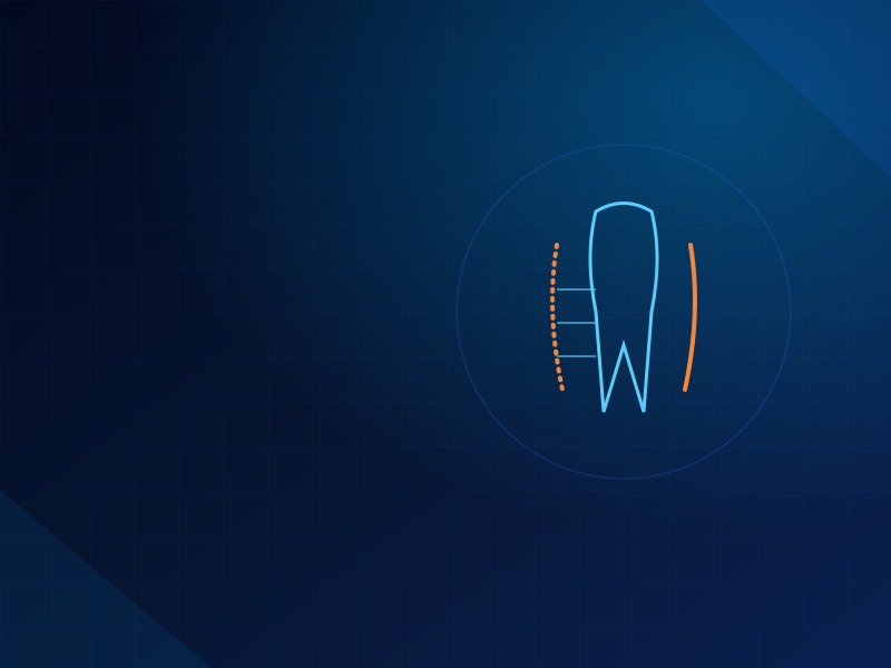
Alveolar Bone Remodeling During Maxillary Incisor Intrusion and Retraction

Alveolar Bone Remodeling During Maxillary Incisor Intrusion and Retraction

Why This Matters

When orthodontists pull upper front teeth back and up (retraction and intrusion) — often to close a gap left after extractions or to correct a "gummy smile" — the surrounding bone doesn't always follow the tooth the way you'd expect. Sometimes it thickens, sometimes it thins, and the outcome is notoriously hard to predict. Unpredictable bone remodeling matters clinically because it can leave visible bone irregularities or gum recession. This study set out to find which initial patient factors actually predict how the bone will respond.

How the Study Was Done

Researchers analyzed before-and-after lateral cephalometric x-rays from 34 patients who had upper incisor retraction and intrusion. They measured bone thickness at three levels below the cementoenamel junction (3mm, 6mm, and 9mm — labeled B1, B2, B3) and tracked the angle between the tooth and the surrounding bone, comparing the initial values to how much each measurement changed after treatment.

Key Findings

  • Bone thickness changes were inconsistent between patients — roughly half showed increased thickness at each level and half showed decreased thickness, even though the group average showed a small statistically significant increase at the 9mm level (B3).
  • That "significant" change was clinically tiny — only about 0.4mm on average — so statistical significance didn't translate into a meaningful clinical difference.
  • Starting tooth/bone angle predicted the outcome: when the incisor or the surrounding bone was more forward-tilted (proclined) to begin with, the bone was more likely to be resorbed (shrink) rather than added during treatment.
  • The amount of retraction itself didn't correlate with bone changes — moving the tooth back further didn't reliably predict more or less bone remodeling.
  • Relative change was linked to starting thickness: patients who started with thinner bone tended to show a larger proportional change, whether gain or loss.
Clinical takeaway: The starting angle between the upper incisor (and its surrounding bone) relative to the Frankfort plane and palatal plane is a meaningful predictor of how the bone will remodel — patients who start more proclined are at higher risk of bone resorption during retraction and intrusion.

What This Means for Practice

Because the direction of bone remodeling can't be fully controlled, the authors recommend carefully assessing these initial angular relationships before treatment — they're one of the few things that actually help predict which patients are more likely to end up with esthetically visible bone irregularities. The study used 2D x-rays, so the authors note that 3D CBCT-based follow-up research would give a clearer, more detailed picture of exactly how the bone contour changes over time.

ORIGINAL SOURCE

Download the full published paper for complete methodology, tables, and references.

Download Full Paper (PDF)

Back to Articles
Blog / Research & Sources
AJODO · 2021

Comparison of Bone-Borne Rapid Maxillary Expansion Effects Between Two Age Groups

Comparison of Bone-Borne Rapid Maxillary Expansion Effects Between Two Age Groups

Why This Matters

Bone-borne palatal expanders — devices anchored entirely to mini-implants rather than to the teeth — are gaining popularity as a way to widen a narrow upper jaw with less unwanted tooth movement (tipping) than traditional tooth-borne expanders. But does patient age at treatment change how well these devices work, both at the bone level and at the tooth level? This study compared outcomes between younger and older patients treated with the same 4-screw bone-borne expander.

How the Study Was Done

Researchers retrospectively studied 24 patients with a narrow upper jaw, split into two groups: age 16 or younger (11 patients) and older than 16 (13 patients). All had a 4-miniscrew bone-borne expander digitally planned and 3D printed insertion guides used for precise miniscrew placement. Before-and-after CBCT scans measured maxillary width, tooth tipping, buccal bone thickness, and how much the expansion at suture level differed from expansion at the tooth level.

Key Findings

  • The device worked in both age groups — 22 of 24 patients (91.7%) achieved actual suture separation, including patients into their late 20s.
  • Skeletal expansion was similar between groups — about 2.8mm in the younger group and 2.1mm in the older group, with no statistically significant difference.
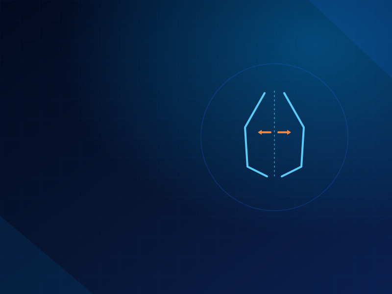
  • Posterior expansion was greater in younger patients (3.54mm vs. 2.84mm), suggesting a more parallel suture opening pattern in the youngest patients compared to a more triangular (front-heavy) opening pattern typically seen with age.
  • Minimal unwanted tooth tipping was seen in both groups — first molar tipping stayed under about 3.6°, notably less than the tipping typically reported with traditional tooth-borne expanders like Hyrax or Haas appliances.
  • Some buccal bone thinning was observed (0.14mm to 0.40mm depending on measurement level), though this did not differ meaningfully between the two age groups.
Clinical takeaway: A fully bone-anchored (4-miniscrew) expander can achieve comparable skeletal expansion with minimal dental side effects in both younger and older patients — making chronological age less of a barrier than it is with conventional tooth-borne expanders.

What This Means for Practice

Digital insertion planning — overlaying an intraoral scan onto the CBCT to plan miniscrew position precisely, then 3D printing surgical guides — helped achieve accurate, bicortical screw placement even given the anatomical constraints of the palate. For older or postpubertal patients who might otherwise be steered toward surgically-assisted expansion, this data supports considering a purely bone-borne approach first, with the caveat that individual suture maturation (not just age) should still be assessed via CBCT before committing to a treatment plan.

ORIGINAL SOURCE

Download the full published paper for complete methodology, tables, and references.

Download Full Paper (PDF)
Back to Articles
Blog / Research & Sources
CASE REPORTS IN DENTISTRY · 2018

Corticopuncture Facilitated Microimplant-Assisted Rapid Palatal Expansion

Corticopuncture Facilitated Microimplant-Assisted Rapid Palatal Expansion

Why This Matters

MARPE usually works well, but there's a real clinical failure mode: sometimes the midpalatal suture is simply too resistant to open, no matter how much force is applied through the jackscrew. This case report documents exactly that scenario in a 35-year-old patient — and demonstrates a minimally invasive fix when MARPE alone isn't enough.

The Case

An adult patient with a narrow upper jaw and a "crooked smile" had a standard 4-miniscrew MARPE device installed. But when she tried to activate it, she couldn't turn the jackscrew — the suture resistance was simply too strong, even with repeated activation attempts over several visits.

Instead of abandoning the nonsurgical approach, the clinicians performed corticopuncture: 8 small perforations (2mm apart, only 5mm deep) drilled directly along the midpalatal suture using a miniscrew as a manual perforating tool, done under local anesthesia as a simple in-office procedure — no surgery, no hospital visit.

What Happened

  • After corticopuncture, the suture opened successfully — the same MARPE device that had been stuck could now be activated normally.
  • CBCT confirmed real skeletal separation: 3.14mm of suture split at the premolar area and 2.06mm at the molar area.
  • Bone actually widened — basal bone width increased by 4.3mm (premolar) and 3.0mm (molar); cortical bone width increased similarly.
  • Dental tipping stayed minimal — averaging just 1.2°, confirming the result was genuinely skeletal rather than just teeth tilting.
  • The suture opened in a "V" shape (more anteriorly than posteriorly) with a parallel split when measured top-to-bottom.
Clinical takeaway: When a MARPE device meets unexpected resistance and won't activate — a real possibility in older or heavily interdigitated patients — corticopuncture along the suture is a minimally invasive way to reduce that resistance without resorting to surgically-assisted expansion (SARPE).

What This Means for Practice

This technique borrows from a broader family of "regional acceleratory phenomenon" methods (corticotomy, micro-osteoperforation, piezocision) that intentionally create controlled bone trauma to accelerate remodeling. The authors note that in this specific case, resistance returned again after 10 more days of activation — suggesting corticopuncture may need to be repeated, or paired with a stiffer expander design, for suture patterns that are especially resistant. Still, as the first documented case of this exact combination, it offers clinicians a practical fallback before jumping straight to surgery when MARPE stalls.

ORIGINAL SOURCE

Download the full published paper for complete methodology, tables, and references.

Download Full Paper (PDF)

Back to Articles
Blog / Research & Sources
PROGRESS IN ORTHODONTICS · 2017

Changes in the Midpalatal and Pterygopalatine Sutures Induced by Micro-Implant-Supported Skeletal Expander

Changes in the Midpalatal and Pterygopalatine Sutures Induced by Micro-Implant-Supported Skeletal Expander

Why This Matters

Most research on palatal expansion focuses on the midpalatal suture — the main line down the center of the palate. But there's another, less-studied structure involved: the pterygopalatine suture, located further back where the maxilla meets the pterygoid processes of the skull base. Some researchers have claimed this suture simply can't be split without surgery. This study used high-resolution 3D imaging to test whether a bone-anchored expander (the Maxillary Skeletal Expander, or MSE) could actually open it.

How the Study Was Done

Researchers analyzed before-and-after CBCT scans from 15 late-adolescent patients (average age 17.2, ranging up to 26) treated with MSE — a 4-miniscrew bone-borne expander. They developed three new 3D reference planes to precisely measure how much the suture opened, whether the opening was symmetrical side-to-side, and whether the deeper pterygopalatine suture also separated.

Key Findings

  • The midpalatal suture opened almost perfectly parallel front-to-back — the separation at the back of the palate (4.3mm) was 90% of the separation at the front (4.8mm). This is very different from traditional tooth-borne expanders, which typically produce a "V-shaped" opening — wide in front, barely open in back.
  • The pterygopalatine suture — the one some researchers thought couldn't be split nonsurgically — actually opened in over half the cases (53% of sutures showed clear separation), something not previously demonstrated with tooth-borne expanders.
  • The split was somewhat asymmetrical side-to-side — on average, one side of the palate moved about 1.1mm more than the other, for reasons not yet fully understood.
  • Patient age and sex didn't meaningfully affect the results — even patients into their mid-20s showed successful, comparable suture separation.
Clinical takeaway: A 4-miniscrew bone-borne expander (MSE) can achieve a more evenly-distributed, parallel expansion of the midpalatal suture than traditional tooth-borne expanders — and can even loosen the pterygopalatine suture further back, something previously thought to require surgery.

What This Means for Practice

Because the expansion force in MSE comes from miniscrews placed well back in the palate (rather than being transmitted through the teeth), it distributes more evenly along the entire suture — explaining the parallel split pattern. This also has downstream implications: the authors note that loosening the pterygopalatine suture could make MSE especially useful as a first step before facemask therapy in patients needing forward maxillary movement, since a looser suture network offers less resistance to that subsequent treatment.

ORIGINAL SOURCE

Download the full published paper for complete methodology, tables, and references.

Download Full Paper (PDF)

Back to Articles
Blog / Research & Sources
IJIRSS · 2025

Digital Design and Fabrication of Direct-Printed Twin Block Appliances

Digital Design and Fabrication of Direct-Printed Twin Block Appliances

Why This Matters

Twin blocks (and their reverse-bite variant) are usually fabricated the old way — pouring casts, hand-bending wires, building acrylic blocks. This paper presents a fully digital, in-house alternative built entirely on free and low-cost software, making a digital twin block workflow accessible to practices without expensive proprietary CAD systems.

Key Findings

  • The entire workflow uses open-source CAD (FreeCAD) paired with a consumer intraoral scanner (Medit i700) — no costly proprietary appliance-design software required.
  • The digital construction bite is captured directly with the scanner, replacing the traditional wax bite registration step.
  • Appliances are designed as splint baseplates with interlocking bite blocks, then 3D printed and post-processed as a single direct-printed piece.
Clinical takeaway: A genuinely low-cost, open-source digital pathway exists for twin block fabrication — useful for practices exploring digital workflows without a large software investment.

ORIGINAL SOURCE

Download the full published paper for complete methodology, tables, and references.

Download Full Paper (PDF)

Back to Articles
Blog / Research & Sources
Australasian Orthodontic Journal · 2025

Digital Workflow to Fabricate a 3D-Printed Monobloc Mandibular Advancement Appliance for Primary Snoring and Obstructive Sleep Apnoea

Digital Workflow to Fabricate a 3D-Printed Monobloc Mandibular Advancement Appliance for Primary Snoring and Obstructive Sleep Apnoea

Why This Matters

Mandibular advancement appliances (MAAs) are a well-established treatment for primary snoring and mild-to-moderate obstructive sleep apnoea, working by holding the lower jaw forward to keep the airway open during sleep. This paper documents a fully digital workflow to design and 3D print a monobloc (single-piece) MAA, extending digital appliance fabrication beyond orthodontics into sleep medicine.

Key Findings

  • The workflow moves from intraoral scan directly to a digitally-designed, single-piece monobloc appliance — skipping lab fabrication steps entirely.
  • A monobloc design (upper and lower fused into one printed piece) simplifies fabrication compared to traditional two-piece adjustable appliances.
  • The digital approach offers a faster, more standardized path to producing MAAs compared to conventional lab-built devices.
Clinical takeaway: The same digital design + 3D printing skillset used for orthodontic appliances extends naturally into airway/sleep appliance fabrication — a useful cross-over application for digitally-equipped practices.

ORIGINAL SOURCE

Download the full published paper for complete methodology, tables, and references.

Download Full Paper (PDF)

Back to Articles
Blog / Research & Sources
2025 · Review

3D Printed Removable Functional Appliances for Early Orthodontic Treatment — Possibilities and Limitations

3D Printed Removable Functional Appliances for Early Orthodontic Treatment — Possibilities and Limitations

Why This Matters

Removable functional appliances (like twin blocks, activators, and bionators) are a mainstay of early Class II treatment. As digital workflows spread, this review looks broadly at where 3D printing genuinely helps — and where it still falls short — for this whole appliance category, not just one specific device.

Key Points

  • 3D printing offers clear advantages in speed, consistency, and reduced lab dependency for functional appliance fabrication.
  • Limitations remain around material properties — printed resins don't always match the flexibility and long-term durability of traditional acrylic.
  • Digital bite registration and appliance activation protocols still need standardization across different printer/material combinations.
Clinical takeaway: 3D printing is a genuine option for functional appliances today, but material selection and activation design still require careful case-by-case judgment rather than a one-size-fits-all digital protocol.

ORIGINAL SOURCE

Download the full published paper for complete methodology, tables, and references.

Download Full Paper (PDF)
Back to Articles
Blog / Research & Sources
Cureus · 2025

Hidden Risks of Miniscrew Assisted Rapid Palatal Expansion (MARPE): A Retrospective Analysis of Treated Cases With Preventive Strategies

Hidden Risks of Miniscrew Assisted Rapid Palatal Expansion (MARPE): A Retrospective Analysis of Treated Cases With Preventive Strategies

Why This Matters

This retrospective review of 24 consecutive MARPE cases (mean age 17.4) looks specifically at complications across two different appliance designs — a 2-screw hybrid MARPE and a 4-screw MSE — with an eye toward practical prevention strategies clinicians can apply.

Key Findings

  • Complications were tracked across both a 2-screw hybrid design (16 cases) and a 4-screw MSE design (8 cases), allowing direct comparison of risk patterns between appliance types.
  • The study emphasizes that most complications are preventable with careful screw diameter/length selection (1.8–2.0mm, 9–11mm) and diligent monitoring.
  • Preventive strategies are proposed based on the specific failure patterns observed in each appliance design group.
Clinical takeaway: Appliance design choice (2-screw vs. 4-screw) meaningfully affects the complication profile — case selection and screw sizing should be tailored accordingly, not treated as interchangeable.

ORIGINAL SOURCE

Download the full published paper for complete methodology, tables, and references.

Download Full Paper (PDF)

Back to Articles
Blog / Research & Sources
Scientific Reports · 2021

Mathematical Modeling of Palatal Suture Pattern Formation: Morphological Differences Between Sagittal and Palatal Sutures

Mathematical Modeling of Palatal Suture Pattern Formation: Morphological Differences Between Sagittal and Palatal Sutures

Why This Matters

The midpalatal suture is a growth center for the maxilla, and its interdigitated (wavy, interlocking) shape is central to how resistant it is to expansion. This basic-science study asks a more fundamental question: what actually determines that wavy pattern in the first place, and how does it differ from other skull sutures like the sagittal suture?

Key Findings

  • The palatal suture is narrower than the sagittal (skull cap) suture, and its interdigitation (waviness) pattern differs measurably in amplitude and geometry.
  • The authors built a mathematical/computational model to explain why these two suture types develop such different shapes despite forming through similar biological processes.
  • This modeling approach helps explain the structural basis for why the palatal suture behaves the way it does under expansion forces.
Why it's relevant clinically: Understanding the biological basis of suture interdigitation helps explain why some patients' palatal sutures resist expansion far more than others — a foundational science piece behind everything MARPE/SARPE clinicians observe at the chair.

ORIGINAL SOURCE

Download the full published paper for complete methodology, tables, and references.

Download Full Paper (PDF)

Back to Articles
Blog / Research & Sources
Case Report · 2025

Maxillary Bone Fracture Due to a Miniscrew-Assisted Rapid Maxillary Expansion: A Case Report

Maxillary Bone Fracture Due to a Miniscrew-Assisted Rapid Maxillary Expansion: A Case Report

Why This Matters

MARPE is generally considered safe, but serious complications — while rare — do happen. This case report documents a maxillary bone fracture in a 32-year-old patient following MARPE activation, offering a sobering, detailed look at what can go wrong and why.

What Happened

  • Five days after activation, the patient developed severe pain and sensory disturbances in the infraorbital region.
  • CT imaging revealed a fracture extending from the infraorbital foramen down to the alveolar process — a genuinely serious structural complication.
  • Symptoms gradually improved over two years, but the psychological impact led the patient to abandon a planned orthognathic surgery altogether.
  • The authors suggest the fracture was related to stress concentration patterns during expansion.
Clinical takeaway: While rare, MARPE-induced fractures are a real risk that deserves a place in informed consent discussions — particularly regarding infraorbital nerve involvement and the potential for lasting psychological impact beyond the physical injury.

ORIGINAL SOURCE

Download the full published paper for complete methodology, tables, and references.

Download Full Paper (PDF)

Back to Articles
Blog / Research & Sources
AJODO · 2013 · Landmark Study

Midpalatal Suture Maturation: Classification Method for Individual Assessment Before Rapid Maxillary Expansion

Midpalatal Suture Maturation: Classification Method for Individual Assessment Before Rapid Maxillary Expansion

Why This Matters

Before this study, there was no standardized way to look at a patient's CBCT and objectively judge how "fused" their midpalatal suture was — a critical factor in deciding whether conventional expansion, MARPE, or surgery is the right call. This paper introduced the classification system that's now widely referenced across the MARPE literature (including several other papers in this library).

Key Findings

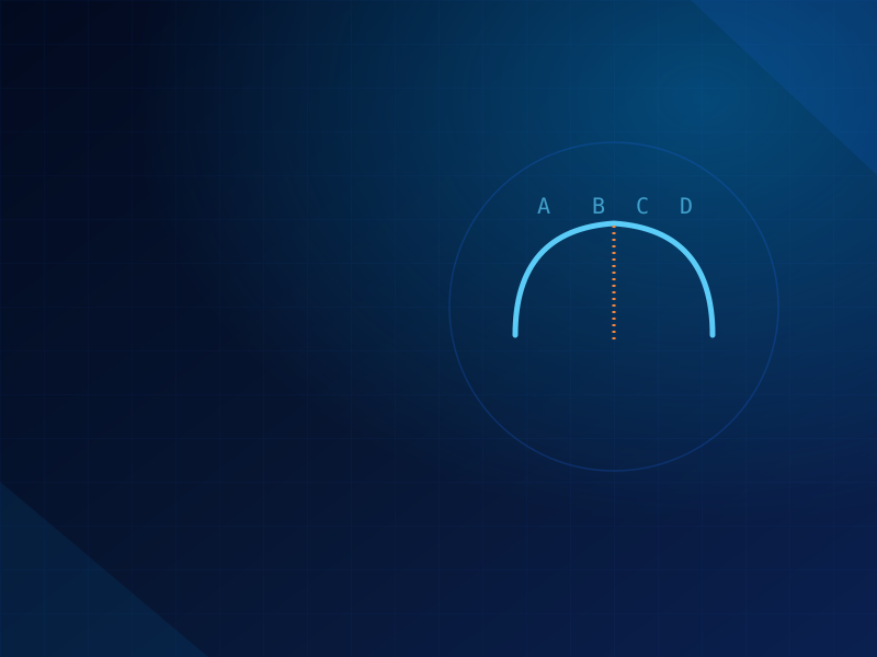
  • The authors examined CBCT scans from 140 subjects (ages 5.6–58.4) to define five distinct maturation stages (A through E) of the midpalatal suture.
  • Stage A is a straight, minimally-interdigitated line (typically young patients); Stage E shows fusion extending anteriorly into the maxilla (typically older, skeletally mature patients).
  • The classification showed strong agreement both within and between examiners, making it reliable enough for routine clinical use.
Clinical takeaway: This 5-stage system (A–E) is the reference standard cited throughout the MARPE literature for deciding treatment approach — essential background for interpreting nearly every other suture-related paper in this library.

ORIGINAL SOURCE

Download the full published paper for complete methodology, tables, and references.

Download Full Paper (PDF)

Back to Articles
Blog / Research & Sources
2021 · Patient Experience Study

Evaluation of Patients' Experiences After Microimplant-Assisted Rapid Palatal Expansion (MARPE) Treatment

Evaluation of Patients' Experiences After Microimplant-Assisted Rapid Palatal Expansion (MARPE) Treatment

Why This Matters

Most MARPE research measures skeletal and dental outcomes on scans — this study instead asks patients directly what the experience was actually like, including pain levels and any adverse effects they noticed, based on interviews with 29 patients (average age 22.8).

Key Findings

  • Clinical photographs, radiographs, and CBCT were combined with a structured patient interview covering pain and adverse effects throughout treatment.
  • Pain scores were tracked specifically during the active activation phase, when force is actively being applied to open the suture.
  • The study captures the patient-reported side of MARPE that chart-review studies (which rely on clinician notes) often miss or under-document.
Clinical takeaway: Patient-reported pain and experience data complements the clinical/radiographic complication data from other studies in this library — useful for setting realistic expectations during informed consent.

ORIGINAL SOURCE

Download the full published paper for complete methodology, tables, and references.

Download Full Paper (PDF)

Back to Articles
Blog / Research & Sources
Seminars in Orthodontics

Principles for Placement and Expansion Protocol for the Bone-First Quadexpander in Adolescents and Adults

Principles for Placement and Expansion Protocol for the Bone-First Quadexpander in Adolescents and Adults

Why This Matters

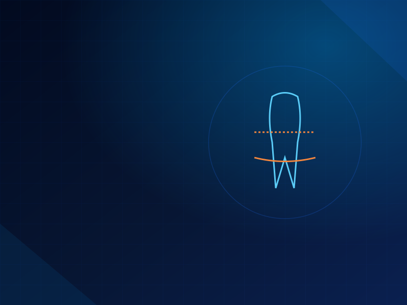
Transverse maxillary deficiency is especially difficult to treat in adults, where the midpalatal and circummaxillary sutures resist conventional tooth-borne expansion. This paper details the placement principles and activation protocol for the "bone-first" Quadexpander — a 4-miniscrew, fully bone-anchored expansion device designed specifically to sidestep those dental side effects.

Key Points

  • The "bone-first" philosophy prioritizes anchoring entirely in bone before any dental engagement, aiming to minimize the buccal tipping and root resorption seen with tooth-borne expanders.
  • Detailed placement principles are given for miniscrew positioning to maximize primary stability in both adolescent and adult palates.
  • The protocol addresses activation sequencing specific to this 4-screw design, distinct from other MARPE/MSE systems.
Clinical takeaway: Device-specific placement protocols matter — the Quadexpander's bone-first design requires its own placement principles rather than assuming techniques from other MARPE systems transfer directly.

ORIGINAL SOURCE

Download the full published paper for complete methodology, tables, and references.

Download Full Paper (PDF)
Back to Articles
Blog / Research & Sources
Orthodontics & Craniofacial Research · 2025

The Effect of the Anteroposterior Position of Miniscrew Assisted Rapid Palatal Expansion on the Expansion Outcomes in Mature Patients

The Effect of the Anteroposterior Position of Miniscrew Assisted Rapid Palatal Expansion on the Expansion Outcomes in Mature Patients

Why This Matters

Where exactly should MARPE miniscrews sit front-to-back in the palate? This study directly tests whether placing the anterior screws more forward vs. more backward relative to the maxilla's center actually changes how much skeletal expansion you get — a question with immediate implications for appliance planning.

Key Findings

  • 32 skeletally mature patients were split into an anterior group (miniscrews ≥5mm forward of the maxilla's center) and a posterior group (≤2mm from or behind center).
  • Before/after CBCT scans measured skeletal, dental, and paramaxillary changes to isolate the effect of screw position specifically.
  • The study proposes a reference point for optimal anteroposterior placement based on the outcomes observed.
Clinical takeaway: Screw position isn't just about avoiding anatomical obstacles — anteroposterior placement measurably influences the skeletal expansion outcome, giving clinicians another concrete lever to optimize treatment planning.

ORIGINAL SOURCE

Download the full published paper for complete methodology, tables, and references.

Download Full Paper (PDF)

Back to Articles
Blog / Research & Sources
Clinical Perspective

The Evolution of the M.A.R.A. Appliance From Analogue to Digital

The Evolution of the M.A.R.A. Appliance From Analogue to Digital

Why This Matters

The M.A.R.A. (Mandibular Anterior Repositioning Appliance) is a fixed functional appliance for Class II correction, invented in 1991 and refined ever since. Drawing on roughly 1,000 collectively treated cases, the authors argue it remains an undervalued, highly versatile Class II option — and walk through how its design has evolved from purely analogue lab work into a digital workflow.

Key Points

  • The M.A.R.A.'s simple, modular components allow individualized adjustments for a wide range of Class II presentations — likened by the authors to a "Swiss army knife" for Class II correction.
  • Digital design and fabrication workflows now allow the same customizability with improved precision and reduced lab turnaround.
  • The paper reflects on two decades-plus of clinical use to argue the appliance deserves more attention in digitally-focused orthodontic practices.
Clinical takeaway: A well-established fixed functional appliance doesn't need to be reinvented to benefit from digital workflows — the M.A.R.A. shows how existing evidence-based appliances can be digitized without losing what made them effective.

ORIGINAL SOURCE

Download the full published paper for complete methodology, tables, and references.

Download Full Paper (PDF)

Back to Articles
Blog / Research & Sources
2025 · Comparative Study

Treatment Outcomes of 3D-Printed Custom and Conventional Mini-Implant Assisted Rapid Palatal Expanders (MARPE)

Treatment Outcomes of 3D-Printed Custom and Conventional Mini-Implant Assisted Rapid Palatal Expanders (MARPE)

Why This Matters

Custom 3D-printed MARPE appliances promise a better anatomical fit than off-the-shelf conventional designs (like the MSE-II) — but does that translate into measurably better expansion outcomes? This retrospective study directly compares the two across 42 patients.

Key Findings

  • 42 patients (ages 16–35) were split evenly into custom 3D-printed (n=21) and conventional MSE-II (n=21) groups, matched by age and sex.
  • Twelve linear distances and four angles were measured on CBCT — including frontozygomatic width, maxillary width, and nasal cavity width — to compare skeletal and dental effectiveness.
  • The comparison isolates whether the added cost/complexity of custom fabrication actually yields a meaningfully different clinical result.
Clinical takeaway: When custom fabrication is being considered as an upgrade over stock MARPE designs, this head-to-head data helps clinicians weigh whether the investment translates into materially better expansion outcomes for their specific patient population.

ORIGINAL SOURCE

Download the full published paper for complete methodology, tables, and references.

Download Full Paper (PDF)
Back to Articles
Blog / Research & Sources
2025 · Case Series

Unwanted Craniofacial Fractures in MARPE-MASPE Patients: A Hidden Risk

Unwanted Craniofacial Fractures in MARPE-MASPE Patients: A Hidden Risk

Why This Matters

This case series takes a hard look at a rare but serious complication: unintended craniofacial fractures occurring during MARPE and MASPE (Miniscrew-Assisted Slow Palatal Expansion) treatment in adults, where the high forces needed to split a mature suture can, in some cases, cause damage beyond the palate itself.

Key Findings

  • Eleven adult clinical cases are presented, all showing unintended craniofacial complications following skeletal midface expansion with miniscrew-assisted devices.
  • The authors discuss the likely biomechanical mechanisms — essentially, the high forces required to overcome a heavily interdigitated suture in skeletally mature patients can transmit stress to unintended areas of the craniofacial skeleton.
  • Both MARPE (rapid) and MASPE (slow) protocols are represented, suggesting the risk isn't limited to one activation speed.
Clinical takeaway: In skeletally mature adults especially, clinicians should watch for signs of resistance during activation and be aware that force doesn't always concentrate exactly where intended — this collection of cases documents where things can go wrong beyond the palate.

ORIGINAL SOURCE

Download the full published paper for complete methodology, tables, and references.

Download Full Paper (PDF)
Back to Articles
Blog / Research & Sources
J. Clin. Med. · Cross-Sectional Study

Effects of Occlusal Contact on Maxillary Alveolar Bone Morphology in Patients With and Without Anterior Open Bite

Effects of Occlusal Contact on Maxillary Alveolar Bone Morphology in Patients With and Without Anterior Open Bite

Why This Matters

Anterior open bite (AOB) means the front teeth never touch when the back teeth are closed together. This study asks a structural question: does the lack of front-tooth contact actually change the shape and thickness of the surrounding alveolar bone compared to patients with normal contact?

Key Findings

  • 50 adults were compared — 25 with AOB and 25 without — measuring alveolar bone crest height and cortical bone thickness (both labial and palatal sides) using CBCT.
  • The comparison targeted both anterior and posterior teeth regions to see whether differences were localized to the open bite area or more generalized.
  • This gives orthodontists concrete anatomical data on what to expect structurally in AOB patients before planning tooth movement in that region.
Clinical takeaway: Understanding baseline bone morphology differences in AOB patients helps set realistic expectations for how much room there actually is to safely move anterior teeth during open bite correction.

ORIGINAL SOURCE

Download the full published paper for complete methodology, tables, and references.

Download Full Paper (PDF)
Back to Articles
Blog / Research & Sources
J. Esthetic & Restorative Dentistry · 2025

Adult Scissor Bite Case Treatment With Moving Teeth Through the Maxillary Sinus

Adult Scissor Bite Case Treatment With Moving Teeth Through the Maxillary Sinus

Why This Matters

Severe scissor bite — where posterior teeth cross so far that normal occlusion is impossible — is a genuinely difficult correction, especially when molars have extruded significantly. This case report documents a 26-year-old patient treated with a technique that intentionally moves teeth through the maxillary sinus region using temporary anchorage devices (TADs).

What Happened

  • Interradicular TADs were used to intrude the extruded molars, control their inclination, and correct the dental midline — the core biomechanical challenges in scissor bite correction.
  • The treatment successfully corrected the scissor bite while achieving functional occlusion and improved facial esthetics.
  • The case demonstrates that TAD-supported tooth movement can safely navigate anatomically complex regions like the maxillary sinus floor when properly planned.
Clinical takeaway: For severe scissor bite with significant molar extrusion, TAD-anchored intrusion — even when it requires moving teeth through sinus-adjacent bone — can be a viable nonsurgical path to a stable, functional correction.

ORIGINAL SOURCE

Download the full published paper for complete methodology, tables, and references.

Download Full Paper (PDF)
Back to Articles
Blog / Research & Sources
Saudi Dental Journal · 2024 · Review

Why Does Maxillary Skeletal Expansion Work With Some Adults and Fail With Others? A Narrative Review

Why Does Maxillary Skeletal Expansion Work With Some Adults and Fail With Others? A Narrative Review

Why This Matters

Every clinician using MARPE has seen it: two adult patients with seemingly similar profiles, and one expands beautifully while the other's suture barely budges. This narrative review pulls together the scattered literature to answer the question directly — what actually separates success from failure in adult maxillary skeletal expansion?

Key Findings

  • Suture maturation stage is the single biggest predictor — patients with more fused sutures (later stages on the Angelieri classification) are significantly more likely to see expansion fail or require surgical assistance.
  • Age alone is an imperfect proxy for suture maturity — chronological age correlates with fusion but with enough individual variability that CBCT assessment remains necessary rather than relying on age cutoffs.
  • Appliance design (screw number, bicortical vs. monocortical engagement) and anchorage quality also meaningfully influence success rates, independent of the patient's underlying anatomy.
  • Circummaxillary sutures beyond the midpalatal suture itself (zygomatic buttresses, pterygomaxillary junctions) contribute additional resistance that isn't always accounted for in treatment planning.
Clinical takeaway: Before starting MARPE in an adult, a CBCT-based suture maturation assessment is far more predictive of success than age or clinical exam alone — this review consolidates the evidence for why that step shouldn't be skipped.

ORIGINAL SOURCE

Download the full published paper for complete methodology, tables, and references.

Download Full Paper (PDF)
Back to Articles
Blog / Research & Sources
UCLA Study · Long-Term Follow-Up

Long-Term Effects of Maxillary Skeletal Expander Treatment on Functional Breathing

Long-Term Effects of Maxillary Skeletal Expander Treatment on Functional Breathing

Why This Matters

MARPE/MSE is often promoted for airway benefits alongside its skeletal effects, but does that improvement in nasal breathing actually last? This study followed 17 patients (average age 19.4) through three checkpoints — before expansion, right after, and after full orthodontic treatment — to see whether breathing gains from MSE hold up over the long term.

Key Findings

  • Breathing was measured both objectively (peak nasal and oral inspiratory flow) and subjectively (patient-reported symptom scores and visual analog scales).
  • Immediately after expansion, all objective breathing measures improved significantly — confirming MSE does open up nasal airflow in the short term.
  • Patient-reported nasal obstruction symptoms also dropped significantly right after expansion, matching the objective airflow improvements.
  • The three-timepoint design (pre-expansion, post-expansion, post-full-treatment) lets the study distinguish immediate effects from those that persist through complete orthodontic treatment.
Clinical takeaway: This adds objective longitudinal evidence to the airway-benefit claims often made about MARPE/MSE — useful for conversations with patients who are motivated by breathing improvement as much as (or more than) bite correction.

ORIGINAL SOURCE

Download the full published paper for complete methodology, tables, and references.

Download Full Paper (PDF)
Back to Articles

Research & Sources

All published research summaries from the Digital Orth Masters library.

Kilbon Appliance Seminars in Orthodontics · 2024 Kilbon Appliance

Kilbon Appliance

2024-05-01 Read More
Assessment of the Bone Thickness of the Palate on CBCT for Placement of Miniscrew-Assisted Rapid Palatal Expansion Appliances AJODO · Research & Sources Palatal Bone Thickness for MARPE

Assessment of the Bone Thickness of the Palate on CBCT for Placement of Miniscrew-Assisted Rapid Palatal Expansion Appliances

2022-06-01 Read More
3D Printed Twin Block: A Feasibility Study Annals of 3D Printed Medicine · 2023 3D Printed Twin Block

3D Printed Twin Block: A Feasibility Study

2023-06-16 Read More
Induced Ankylosis of a Primary Molar for Skeletal Anchorage as an Alternative to Mini-Implants Progress in Orthodontics · 2015 Induced Ankylosis Anchorage

Induced Ankylosis of a Primary Molar for Skeletal Anchorage as an Alternative to Mini-Implants

2015-06-18 Read More
A Retrospective Analysis of the Complications Associated with Miniscrew-Assisted Rapid Palatal Expansion AJO-DO Clinical Companion · 2022 MARPE Complications

A Retrospective Analysis of the Complications Associated with Miniscrew-Assisted Rapid Palatal Expansion

2022-10-01 Read More
Alveolar Bone Remodeling During Maxillary Incisor Intrusion and Retraction Progress in Orthodontics · 2019 Alveolar Bone Remodeling

Alveolar Bone Remodeling During Maxillary Incisor Intrusion and Retraction

2019-12-23 Read More
Comparison of Bone-Borne Rapid Maxillary Expansion Effects Between Two Age Groups AJODO · 2021 Bone-Borne Expansion by Age

Comparison of Bone-Borne Rapid Maxillary Expansion Effects Between Two Age Groups

2021-01-01 Read More
Corticopuncture Facilitated MARPE Case Reports in Dentistry · 2018 Corticopuncture + MARPE

Corticopuncture Facilitated Microimplant-Assisted Rapid Palatal Expansion

2018-12-06 Read More
Pterygopalatine Suture Changes Progress in Orthodontics · 2017 Pterygopalatine Suture Changes

Changes in the Midpalatal and Pterygopalatine Sutures Induced by Micro-Implant-Supported Skeletal Expander

2017-11-01 Read More
Digital Design and Fabrication of Direct-Printed Twin Block Appliances IJIRSS · 2025 Digital Twin Block Workflow

Digital Design and Fabrication of Direct-Printed Twin Block Appliances

2025-01-01 Read More
Digital Workflow to Fabricate a 3D-Printed Monobloc Mandibular Advancement Appliance for Primary Snoring and Obstructive Sleep Apnoea Australasian Orthodontic Journal · 2025 Digital Sleep Apnoea Appliance

Digital Workflow to Fabricate a 3D-Printed Monobloc Mandibular Advancement Appliance for Primary Snoring and Obstructive Sleep Apnoea

2025-01-01 Read More
3D Printed Removable Functional Appliances for Early Orthodontic Treatment — Possibilities and Limitations Review · 2025 3D Printed Functional Appliances

3D Printed Removable Functional Appliances for Early Orthodontic Treatment — Possibilities and Limitations

2025-01-01 Read More
Hidden Risks of Miniscrew Assisted Rapid Palatal Expansion (MARPE): A Retrospective Analysis of Treated Cases With Preventive Strategies Cureus · 2025 MARPE Hidden Risks

Hidden Risks of Miniscrew Assisted Rapid Palatal Expansion (MARPE): A Retrospective Analysis of Treated Cases With Preventive Strategies

2025-09-25 Read More
Mathematical Modeling of Palatal Suture Pattern Formation: Morphological Differences Between Sagittal and Palatal Sutures Scientific Reports · 2021 Suture Pattern Modeling

Mathematical Modeling of Palatal Suture Pattern Formation: Morphological Differences Between Sagittal and Palatal Sutures

2021-04-27 Read More
Maxillary Bone Fracture Due to a Miniscrew-Assisted Rapid Maxillary Expansion: A Case Report J Clin Med · 2025 MARPE Fracture Case

Maxillary Bone Fracture Due to a Miniscrew-Assisted Rapid Maxillary Expansion: A Case Report

2025-03-13 Read More
Midpalatal Suture Maturation: Classification Method for Individual Assessment Before Rapid Maxillary Expansion AJODO · 2013 Suture Maturation Stages

Midpalatal Suture Maturation: Classification Method for Individual Assessment Before Rapid Maxillary Expansion

2013-11-01 Read More
Evaluation of Patients' Experiences After Microimplant-Assisted Rapid Palatal Expansion (MARPE) Treatment Patient Experience · 2021 MARPE Patient Experience

Evaluation of Patients' Experiences After Microimplant-Assisted Rapid Palatal Expansion (MARPE) Treatment

2021-01-01 Read More
Principles for Placement and Expansion Protocol for the Bone-First Quadexpander in Adolescents and Adults Seminars in Orthodontics Quadexpander Protocol

Principles for Placement and Expansion Protocol for the Bone-First Quadexpander in Adolescents and Adults

2024-01-01 Read More
The Effect of the Anteroposterior Position of Miniscrew Assisted Rapid Palatal Expansion on the Expansion Outcomes in Mature Patients Ortho & Craniofacial Research · 2025 MARPE Screw Position

The Effect of the Anteroposterior Position of Miniscrew Assisted Rapid Palatal Expansion on the Expansion Outcomes in Mature Patients

2025-01-01 Read More
The Evolution of the M.A.R.A. Appliance From Analogue to Digital Clinical Perspective M.A.R.A. Goes Digital

The Evolution of the M.A.R.A. Appliance From Analogue to Digital

2024-01-01 Read More
Treatment Outcomes of 3D-Printed Custom and Conventional Mini-Implant Assisted Rapid Palatal Expanders (MARPE) Comparative Study · 2025 Custom vs. Conventional MARPE

Treatment Outcomes of 3D-Printed Custom and Conventional Mini-Implant Assisted Rapid Palatal Expanders (MARPE)

2025-01-01 Read More
Unwanted Craniofacial Fractures in MARPE-MASPE Patients: A Hidden Risk Case Series · 2025 Craniofacial Fracture Risk

Unwanted Craniofacial Fractures in MARPE-MASPE Patients: A Hidden Risk

2025-01-01 Read More
Effects of Occlusal Contact on Maxillary Alveolar Bone Morphology in Patients With and Without Anterior Open Bite J Clin Med Open Bite Bone Morphology

Effects of Occlusal Contact on Maxillary Alveolar Bone Morphology in Patients With and Without Anterior Open Bite

2024-01-01 Read More
Adult Scissor Bite Case Treatment With Moving Teeth Through the Maxillary Sinus Clinical Case · 2025 Scissor Bite via Sinus

Adult Scissor Bite Case Treatment With Moving Teeth Through the Maxillary Sinus

2025-01-01 Read More
Why Does Maxillary Skeletal Expansion Work With Some Adults and Fail With Others? A Narrative Review Saudi Dental Journal · 2024 Why MARPE Fails

Why Does Maxillary Skeletal Expansion Work With Some Adults and Fail With Others? A Narrative Review

2024-04-26 Read More
Long-Term Effects of Maxillary Skeletal Expander Treatment on Functional Breathing Long-Term Follow-Up MSE & Breathing

Long-Term Effects of Maxillary Skeletal Expander Treatment on Functional Breathing

2024-01-01 Read More